Exploring the Global State Space of the Brain
Combining network-wide microwire constellations with active optogenetic probing to understand brain state and seizure risk.
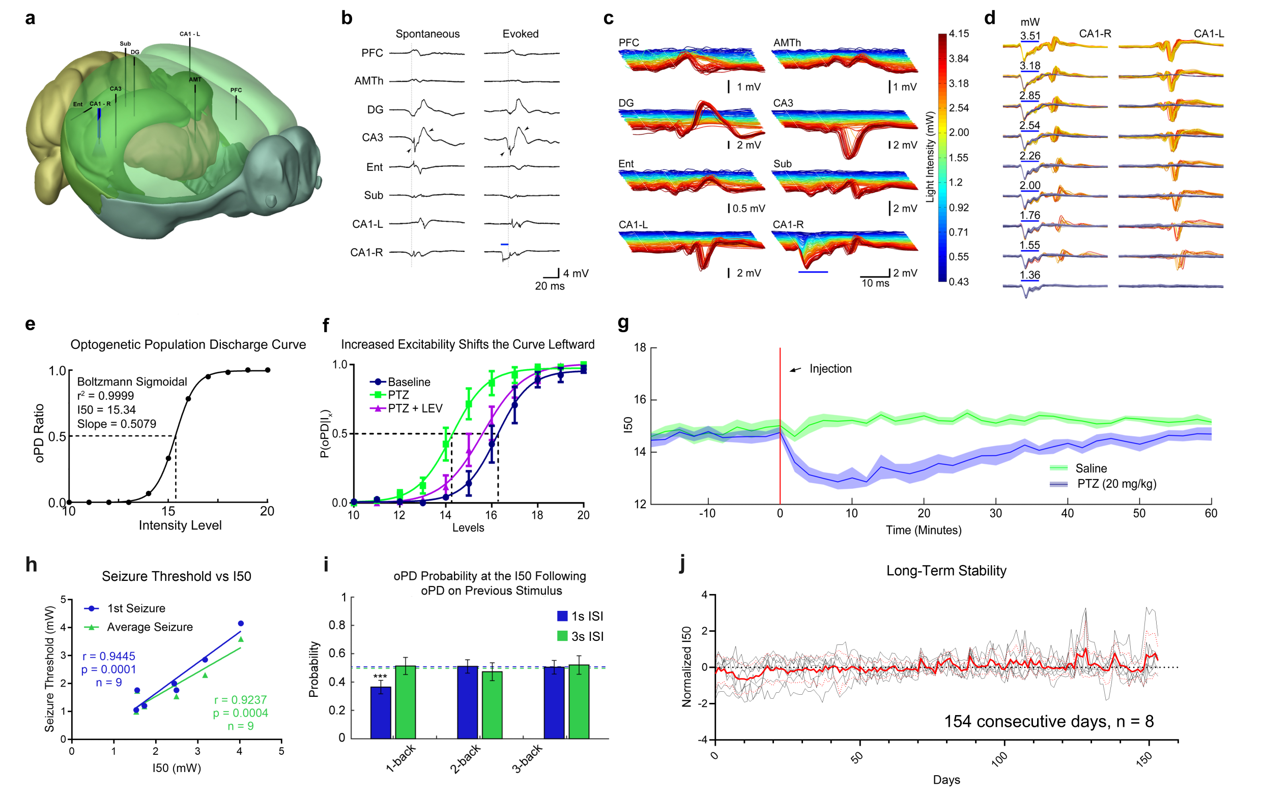
Adapted from Klorig et. al. 2019
Measuring Seizure Risk
We’ve developed a novel method, the optogenetic population discharge threshold, which provides a measure of seizure risk over time. Using this method, we are attempting to understand the factors which govern seizure risk on timescales ranging from individual cycles of sleep oscillations to the multidian oscillations that modulate risk over weeks and months.
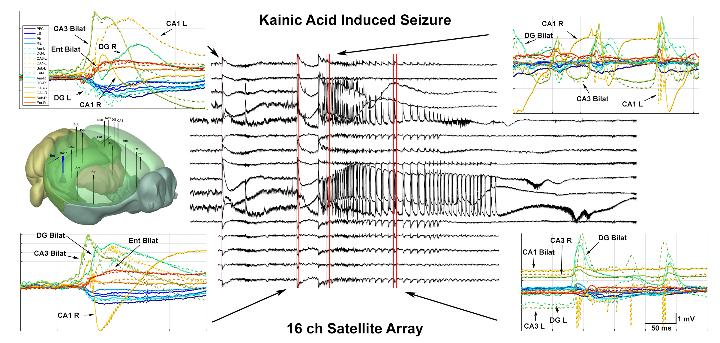
Seizure Forecasting
Using long-term longitudinal datasets we have begun the process of mapping seizure risk across brain states. Using our novel techniques, we have identified high and low risk periods. Current work is focused on understanding the underlying factors which govern seizure risk.
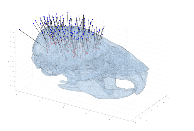
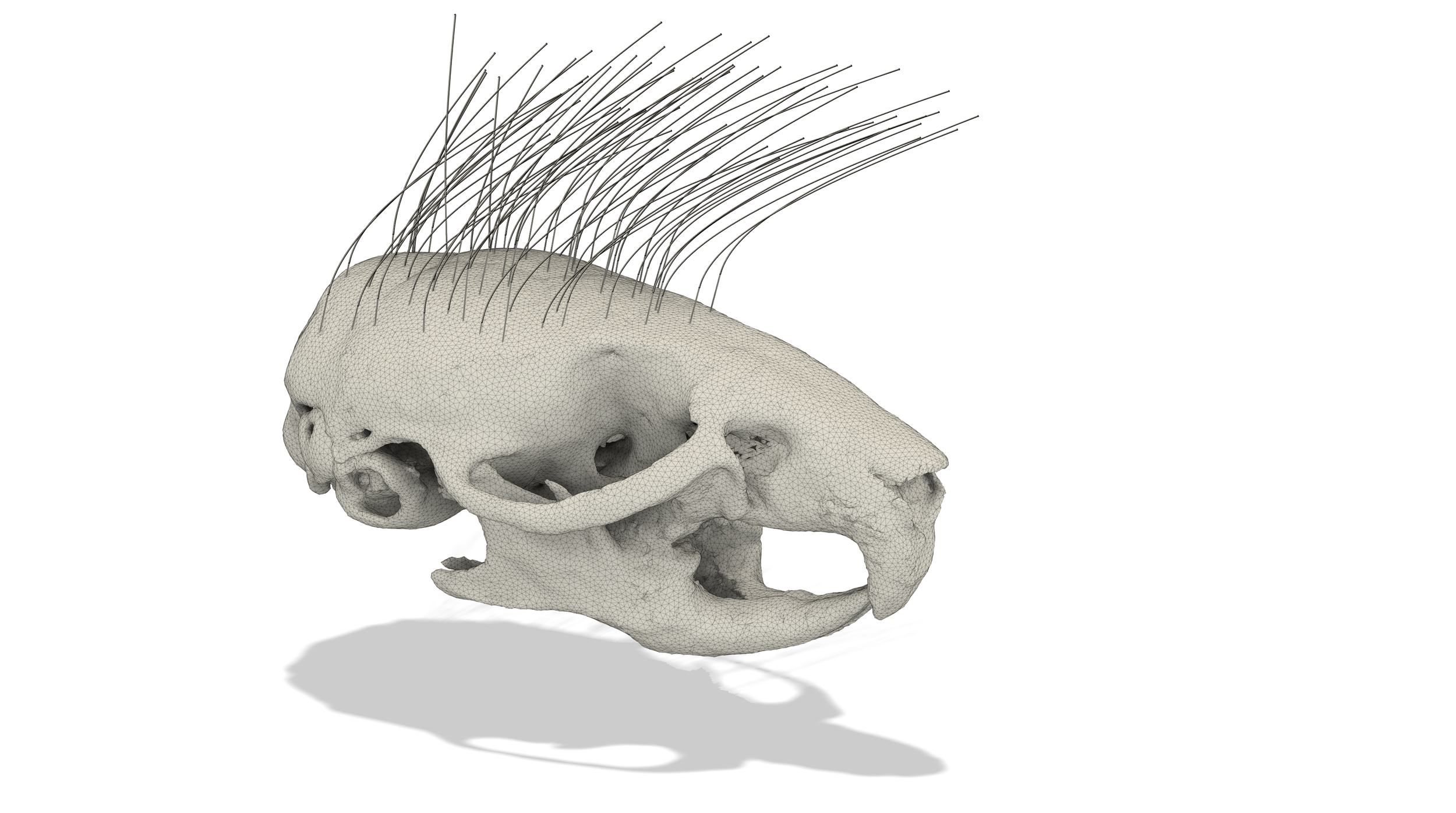
Automated Microsurgery
We are actively developing automated techniques for minimally invasive placement of dispersed microwire constellations for whole brain depth recording.